Lower Back And Leg Muscle Diagram / Muscles Move And Support The Spine - Our latest youtube film is ready to run.. The bones of the pelvis and lower back work together to support the body's weight, anchor the abdominal and hip muscles, and protect the delicate vital organs of the vertebral and abdominopelvic cavities. Related posts of muscles and tendons of the leg muscle anatomy labeling. While factors like what your pain feels like—stabbing, burning, or cramping, and so on—can provide insight, oftentimes, a detailed physical examination and/or an imaging test are needed to clinch the diagnosis. This is a small muscle in the back of the lower leg. English french german latin spanish view all.
Posterior compartment, also known as the flexor compartment; These structures work together to support the body, enable a range of movements, and send messages from the brain to. Below you'll see diagrams along with the names of the back muscles that may be the cause of. Extends spine and trunk back. Write down the muscles of the thigh in the table below and, for each, give the location of that muscle and what effect contracting that muscle has.
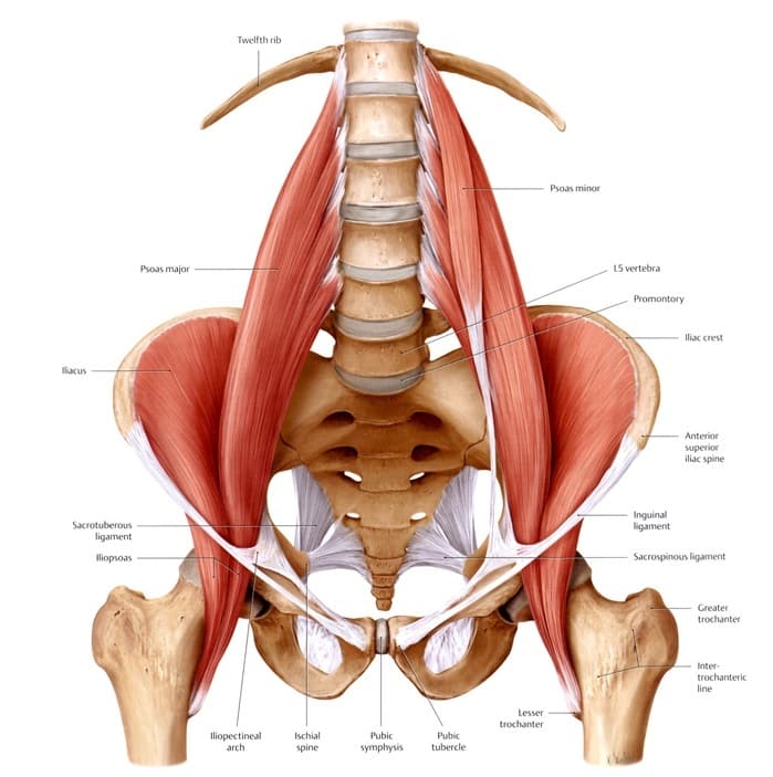
Leg Definition Bones Muscles Facts Britannica from cdn.britannica.com Surprisingly to many, trigger points in the soleus can be the origin of tight hamstrings and lower back pain. Likewise, there are muscles in other parts of the body that help support and move the spine. The bones of the pelvis and lower back work together to support the body's weight, anchor the abdominal and hip muscles, and protect the delicate vital organs of the vertebral and abdominopelvic cavities. The lower leg is a major anatomical part of the skeletal system. It lies between the knee and the ankle, while the upper leg lies between. Other muscles beyond the back also help move the head, shoulders, arms, and legs. Piriformis muscle anatomy ultrasound 12 photos of the piriformis muscle anatomy ultrasound piriformis muscle anatomy ultrasound, human muscles, piriformis muscle anatomy ultrasound. Observe the leg muscle diagram posted above and notice that there are many parts in the muscles.the largest muscle masses in the leg are present in the thigh and the calf.
While a pulled muscle in your lower back could potentially cause a pinched nerve, this can also be caused by a herniated disc in your spine.
Muscle anatomy spandex 12 photos of the muscle anatomy spandex muscle anatomy spandex, human muscles, muscle anatomy spandex Lower leg pain is common, but it can be tricky sorting out its many potential causes. There are many muscles located in the lower leg, but there are three that are particularly well known—the gastrocnemius and the soleus, which are the most powerful muscles in the lower leg, and the anterior tibialis. The following diagram illustrates the actions of the terms adduction, abduction, flexion and extension at the different joints. Other muscles beyond the back also help move the head, shoulders, arms, and legs. The thigh (proximal lower limb) muscles are arranged into three compartments : Shoulder muscle anatomy neck muscle anatomy anatomy back shoulder muscles gross anatomy chest muscles core muscles human anatomy drawing human body anatomy. 69 inches (5′ 9″) long. For more anatomy content please follow us and visit our website: This is a small muscle in the back of the lower leg. It also decelerates dorsi flexion of the foot. Related posts of lower leg muscles diagram muscle anatomy spandex. A reduction in pain, stiffness and muscle tightness is noticeable after a few treatments, often a difference is felt after the first treatment.
It also decelerates dorsi flexion of the foot. Hamstrings (made of 3 muscles): It also supplies sensation to the sole of the foot, the ankle, the. The achilles tendon is also located in the lower leg. Likewise, there are muscles in other parts of the body that help support and move the spine.
Anatomy Of The Lower Back Elliot S Site from elliottelford.com For more anatomy content please follow us and visit our website: Medial compartment, also known as adductor compartment; The back consists of the spine, spinal cord, muscles, ligaments, and nerves. More articles about back pain Anterior compartment, also known as the extensor compartment; The lower part of the trapezius ascends and depresses the scapula, while the transverse or middle region of the trapezius is what retracts the. Hamstrings (made of 3 muscles): There are many muscles located in the lower leg, but there are three that are particularly well known—the gastrocnemius and the soleus, which are the most powerful muscles in the lower leg, and the anterior tibialis.
The sciatic nerve primarily supplies the muscles of the lower leg, including the calf, ankle, and the back portion of the knee.
Related posts of muscles of the lower back and hip diagram piriformis muscle anatomy ultrasound. This is a small muscle in the back of the lower leg. 69 inches (5′ 9″) long. A basic understanding of the anatomy of your lower back can help you identify and differentiate a problem that commonly affects this region, such as localized muscle pain or sciatica. Anterior compartment, also known as the extensor compartment; For example, some muscles located in the chest also help move the shoulders. There are many muscles located in the lower leg, but there are three that are particularly well known—the gastrocnemius and the soleus, which are the most powerful muscles in the lower leg, and the anterior tibialis. It also decelerates dorsi flexion of the foot. There are three parts to the trapezius. We are pleased to provide you with the picture named muscles of lower back diagram.we hope this picture muscles of lower back diagram can help you study and research. The thigh (proximal lower limb) muscles are arranged into three compartments : Shoulder muscle anatomy neck muscle anatomy anatomy back shoulder muscles gross anatomy chest muscles core muscles human anatomy drawing human body anatomy. Extends spine and trunk back.
Observe the leg muscle diagram posted above and notice that there are many parts in the muscles.the largest muscle masses in the leg are present in the thigh and the calf. While factors like what your pain feels like—stabbing, burning, or cramping, and so on—can provide insight, oftentimes, a detailed physical examination and/or an imaging test are needed to clinch the diagnosis. This is a small muscle in the back of the lower leg. The bones of the pelvis and lower back work together to support the body's weight, anchor the abdominal and hip muscles, and protect the delicate vital organs of the vertebral and abdominopelvic cavities. Our latest youtube film is ready to run.
Arthritis In The Back Symptoms Types Of Back Arthritis Treatment from creakyjoints.org Anterior compartment, also known as the extensor compartment; Upon exiting the vertebral canal, the spinal nerves of the lower back form into two networks known as the lumbar and sacral plexuses. The trapezius or trapezoid muscles are two paired muscles that extend from the base of the thoracic vertebrae in the spine to the occipital bone and run out to the spine of the scapula. These structures work together to support the body, enable a range of movements, and send messages from the brain to. While a pulled muscle in your lower back could potentially cause a pinched nerve, this can also be caused by a herniated disc in your spine. Knowledge of the structures in your lumbar spine can also help you communicate with your doctor about lower back problems. For more anatomy content please follow us and visit our website: A basic understanding of the anatomy of your lower back can help you identify and differentiate a problem that commonly affects this region, such as localized muscle pain or sciatica.
The lumbar plexus supplies nerves to the skin and muscles of the lateral abdominal region, thigh, anterior thigh, and external genitals.
The lower leg is a major anatomical part of the skeletal system. Posterior compartment, also known as the flexor compartment; Shoulder muscle anatomy neck muscle anatomy anatomy back shoulder muscles gross anatomy chest muscles core muscles human anatomy drawing human body anatomy. Your leg muscles are some of the hardest working muscles in your body. Upon exiting the vertebral canal, the spinal nerves of the lower back form into two networks known as the lumbar and sacral plexuses. Hamstrings (made of 3 muscles): The femoral, saphenous, obturator, and lateral femoral cutaneous nerves all extend from the lumbar plexus into the muscles and skin of the thigh and leg. Anterior compartment, also known as the extensor compartment; The back consists of the spine, spinal cord, muscles, ligaments, and nerves. Pain in your calf or thigh can be caused by muscle cramps, a pulled or strained muscle, or issues related to your nerves. Related posts of muscles of the lower back and hip diagram piriformis muscle anatomy ultrasound. Muscle anatomy spandex 12 photos of the muscle anatomy spandex muscle anatomy spandex, human muscles, muscle anatomy spandex A reduction in pain, stiffness and muscle tightness is noticeable after a few treatments, often a difference is felt after the first treatment.

0 Comments:
Posting Komentar